Capitalizing on the Heart of the Action
LETTER FROM THE DEPARTMENT CHAIR
Our graduates are making a difference! The culture of the BME Department is inclusive, energetic, intellectually challenging, and inviting. Coming to us from all over the world, BME students join an engaging community that provides numerous opportunities for career and leadership skill development as well as community outreach and involvement with BME professional societies. BME faculty conduct a wide range of biomedical research across many disciplines of engineering and medicine. We receive prestigious research grants from the National Institutes of Health, National Science Foundation, Department of Defense, American Heart Association, private foundations, and industry. BME graduate students, postdoctoral scientists, and undergraduate students are the essential component of the faculty’s research initiatives and are critical to the Department’s mission to train the next generation of biomedical engineers.
GW’s location in the heart of Washington, D.C., provides BME faculty and students with a wealth of research, internship, and cultural opportunities. The Department is located just across the street from the GW School of Medicine and Health Sciences and GW Hospital, which provide stimulating and collaborative environments for cross disciplinary education and research. The university also is a few blocks from the White House, Washington Monument, Lincoln Memorial, Smithsonian National Museums, and many other national landmarks. We are close, physically and professionally, to numerous federal government institutions and research laboratories, including the NIH, the U.S. Patent and Trademark Office, and the FDA.
BME students are thus regularly exposed to a rich variety of research and internship opportunities and scientific interactions that are not available at most other universities. With internationally renowned faculty, state-of-the-art research labs, and unique academic programs and opportunities that stem from our location, the Department of Biomedical Engineering offers a one-of-a-kind education that opens doors for our students.
Murray H. Loew, Ph.D., P.E.
Professor and Chair,
Department of Biomedical Engineering
ASSISTIVE ROBOTICS AND TELE-MEDICINE (ART-MED) LAB
The Assistive Robotics and Tele-Medicine (ART-Med) Lab is directed by Professor Chung Hyuk Park with a focus on developing humanized intelligence through learning human behaviors and knowledge. The main research areas of ARTMed lab are Assistive Robotics and Healthcare robotics, with specialization in (1) social robots for children with autism, (2) multi-modal perception and mutual learning in human-robot interaction (HRI), (3) digital health and artificial intelligence (AI), and (4) telemedical robotics. Through these research topics, Prof. Park and the members of ARTMed lab aim to have societal impact and increase quality of life
through their intelligent robotic systems and advanced AI algorithms. ART-Med lab has been funded by NIH, NSF, CTSI-CN, KIAT, and GW OVPR, with doctoral and master’s students, several undergraduate researchers and research interns, among whom more than 60% are female students or students from under-represented groups. Companion courses (Introduction to Assistive Robotics, and Socially Assistive Robotics and Interactive Learning) are offered to provide hands-on learning. The collaborators of ART-Med lab are affiliated with the Johns Hopkins University, Georgia Institute of Technology, Virginia Tech., Kennedy Krieger Institute, Southern Methodist University,
Rochester Institute of Technology, and Korea University. The long-term goal of research in ART-Med lab is to have real impact on healthcare and quality of life in our daily living through smart and interactive robotic assistance and specialized AI support.
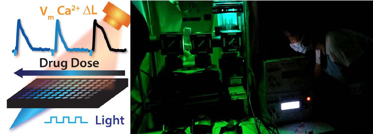
USING LIGHT AND HUMAN STEM-CELL DERIVED HEART CELLS TO ADVANCE PERSONALIZED MEDICINE
Professor Emilia Entcheva’s laboratory (the Cardiac Optogenetics & Optical Imaging Laboratory) combines biophotonics tools with human stem-cell-derived cardiomyocyte technology and gene editing approaches to aid the advancement of personalized medicine.
Personal responses to therapeutics vary based on our genetic background and other markers that make us different. Personalized medicine aims to tailor drug development to individuals, ideally using their own cells for testing. The goal is to maximize efficacy and minimize undesired side effects (e.g., a smart selection of an anti-cancer treatment on individual basis can reduce cardiotoxicity effects). Since 2007, scientists have developed practically noninvasive methods to harvest patient’s cells using blood, urine or skin samples and to turn them into powerful stem cells with the potential to then generate in a dish heart cells, neurons and other types carrying the genetic signature of the patient.
Our laboratory uses these human stem-cell-derived heart cells to engineer live heart tissues and use them for drug discovery and drug testing. To develop commercially viable platforms for drug testing using these personalized heart models in a dish, the lab integrates optical stimulation and optical recording by leveraging optogenetics - the use of optics, lightsensitive proteins and genetic engineering to control and monitor cellular responses in a highly parallel manner. This translational approach is poised to accelerate the optimization of stem-cell-derived heart cells not only for drug testing but also for future regenerative applications, i.e., to repair damaged heart tissue.
We have knowhow and intellectual property in this area and have pioneered the “all-optical cardiac electrophysiology” techniques, especially as applied to human stem-cell-derived cardiomyocytes. Our work is funded by the National Science Foundation and the National Institutes of Health. Beyond the drug testing applications, our technology can be applied to the whole heart to stimulate by light and pave the way to new optical cardiac devices, such as smart optical pacemakers and defibrillators. The timeframe for translation of these devices is substantially longer than the one for adopting our personalized medicine platforms.
THE KAY LAB OF CARDIAC RESEARCH
Heart failure affects nearly 23 million people worldwide and its prevalence is projected to increase more than 40% in the next 15 years. A hallmark of heart failure is elevated cardiac sympathetic activity and parasympathetic withdrawal, an imbalance of the autonomic nervous system that contributes to cardiac dysfunction, inflammation, and arrhythmias. Recent work to improve heart failure treatments has invigorated interest in restoring balance within the autonomic nervous system, particularly in elevating parasympathetic activity to the heart.
With a focus on neurocardiology, the interaction between the brain and heart, Professor Matt Kay’s research is shedding new light on the important role of the cardiac parasympathetic system during health and disease. Prof. Kay and his team tackle important unanswered questions that involve heart failure and its comorbidities: myocardial infarction and sleep apnea; questions that can only be answered by rigorous engineering approaches and multidisciplinary collaborations.
Their work is largely integrative, where cardiovascular disease and therapies are studied in-vivo using echocardiography and telemetry, ex-vivo using high speed optical imaging, and at the subcellular level using protein assays, scanning electron microscopy, and transcriptomics.
With instrumentation developed in the lab, Prof. Kay’s trainees study living hearts using advanced optical mapping of electromechanics and metabolism, time resolved absorbance spectroscopy, optogenetics, and chemogenetics. Cardiac arrhythmias are studied using panoramic optical mapping of membrane potential. NADH imaging provides insight into metabolic fluctuations during myocardial ischemia and reperfusion. High-speed optical spectroscopy quantifies intracellular alterations of myoglobin oxygenation and mitochondrial redox state.
Recent accomplishments of Prof. Kay’s team include developing and testing new approaches for acute and chronic control of the cardiac neuronal network to study and prevent the development of cardiac dysfunction and arrhythmias during cardiovascular disease. These achievements contribute to the design of new clinical devices and pharmaceutical therapies to prevent sudden cardiac death and to reduce the debilitating impact of heart failure and its comorbidities. In all their projects, the Kay Lab team enjoys valuable multidisciplinary collaborations with outstanding investigators within the BME Department, the GW School of Medicine and Health Sciences, the GW Medical Faculty Associates, and beyond GW.
ENGINEERING PERSONALIZED DEVICES
Professor Luyao Lu’s lab explores the next-generation soft, lightweight, biocompatible materials and devices such as transparent microelectrodes, photodetectors, solar cells, and light-emitting diodes. Our short-term goal is to provide advanced electrical and optical microsystems that can seamlessly integrate with and investigate the dynamics of soft biological systems at the micro/nano scales. Our long-term goal is to develop bio-integrated electronics that can facilitate health monitoring, personalized medicine design, organs-on-chips development, and accurate disease diagnosis. Our research is supported by external funding sources such as NSF and NIH as well as multiple GW internal funds. To accomplish our multidisciplinary research goals, we collaborate with research groups from both inside GW and other institutions. Below are highlights of some current research topics in the Lu lab:
-
Develop high-performance miniaturized wireless implants that can provide advanced capabilities in high spatio-temporal resolution controlling and monitoring of cell/ tissue function in freely behaving test subjects. For example, we developed battery-free, fully implantable, flexible wireless photometers to continuously record brain activity in small rodent models over time in complex 3D environments in a way not possible with existing technologies.
-
Design optically transparent microelectrodes with excellent biocompatibility, biochemical stability, and/or bioresorbablility to allow acute or chronic simultaneous coupling of time-resolved electrophysiology data with optically measured, spatially-resolved cell activity. In addition, we investigate scalable fabrication techniques to integrate those materials into soft large-area wafer-scale microelectrode arrays for conformal interfacing with curved tissue/organ systems.
-
Develop multifunctional optoelectronics systems to combine electrophysiology with complementary genetically targeted optical methods for investigating cellular processes under programmed modulation with cell specificity. Those tools are crucial to studying the causal connectivity and function of various biological systems such as the brain and the heart.
-
Design effective wireless, flexible, and self-powering devices to support continuous uninterrupted operations of various wearable and implantable biomedical systems. Those devices will integrate energy harvesting components such as solar cells with energy storage components including batteries and supercapacitors into soft and biocompatible formats.
NOVEL DESIGNS FOR TARGETING AND ELIMINATING CANCER CELLS
Professor Anne Papa’s lab focuses on applying bioengineering principles to develop cell-based therapeutic and diagnostic tools for biomedical applications, in the fields of metastatic cancer and vascular disorders.
Their work in metastatic cancer has been centered on a better understanding of the dynamics and interactions of metastasizing cancer cells in the circulation. Such metastatic cancer cells that have leaked from the original site of the tumor into the bloodstream, can lead to the formation of new distant sites of disease and profoundly influence success of cancer treatments and patient outcomes. Developing detailed knowledge of cancer cells in the circulation has been instrumental in engineering novel designs for targeting and eliminating cancer cells in the blood, as well as capturing those cells for further analysis. For example, the lab currently uses platelets as a means of targeting circulating tumor cells due to their high and natural bioaffinity with cancer cells. The goal is to load platelets with various drugs to both target and modulate circulating tumor cell fate and survival.
The lab also studies how cancer cell-derived exosomes influence platelet function and may contribute to cancer-associated thrombosis, a leading cause of mortality and morbidity for cancer patients. Other areas of work in the lab include studies of how nanoparticles influence essential blood clotting and cellular functions, as well as developing novel targeted therapies for acute emergency vascular disorders due to blood clots, such as in ischemic strokes, heart attacks and pulmonary embolism.
Master of Engineering in Medical Device Engineering and Policy
A unique online program that awards a Master of Engineering in regulatory biomedical engineering by providing students with the understanding of the engineering and regulatory basis of medical device safety and effectiveness. Courses in biomedical engineering and regulatory affairs provide students with the set of engineering skills needed for medical device development, evaluation and commercialization. This degree requires 30 credits in 10 online courses, designed for working students.
Learn more at getinfo.seas.gwu.edu/rbme


